eKonsey
Medical Consulting

Whatsapp
+90 549 804 0308

e-mail
dr@tibeterdogru.com

URETERAL STRICTURE

What is Ureter?

Urinary canal that carries the urine produced in the kidney to the bladder is called ureter. Ureter starts from the right and left kidneys, continues to the bladder, and reaches at the bladder in the area called the trigone.

The region where the ureter goes out from the kidney is called UPJ in short and the other region where it connects to the bladder is called UVJ.

The length of the ureter is between 20 and 25 cm for adults. The inner diameter of the canal varies between 6 and 8 mm. The region where iliac vessels pass outside the UPJ and UVJ is the narrowest areas of the inner diameter of the ureter.

What are the Causes of Ureteral Stenosis?

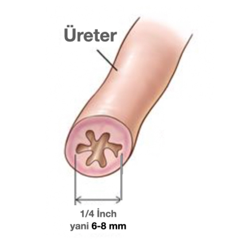
Ureteral canals are very delicate and are supplied by capillaries. These delicate canals can be easily damaged during endoscopic surgery, open surgery or by external trauma.

Laser and endoscopic devices used for interventions in the stones falling into the ureter may cause ureteral injury, and especially following the intestinal and gynecological surgeries, some other injuries or cuts may also occur in the ureters.
In the cases of ureteral stenosis, if the length of the stricture is less than 2 cm, there may be a chance of opening it endoscopically. However, especially if renal functions are affected and ureteral nutrition is impaired in the area where the stenosis exists, opening the stenosis with endoscopic treatment does not help and the stenosis recurs in a short time.
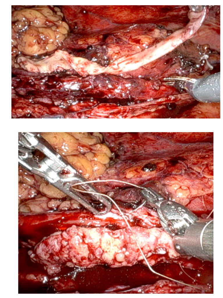
Therefore, in such cases, the narrow part of the ureter is removed, and the canal is reconnected to itself. This is called ureteroplasty end-to-end anastomosis.

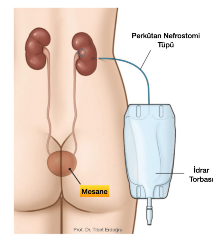
However, in some ureteral injuries, the stenosis may be longer than 2 cm and the nutrition and blood supply in this narrow part of the ureter are impaired. If the narrow part is removed, the intact ureteral ends are not brought closer together, which poses a significant problem. Until today, patients with such cases usually have had to live with nephrostomy tubes inserted from the waist.

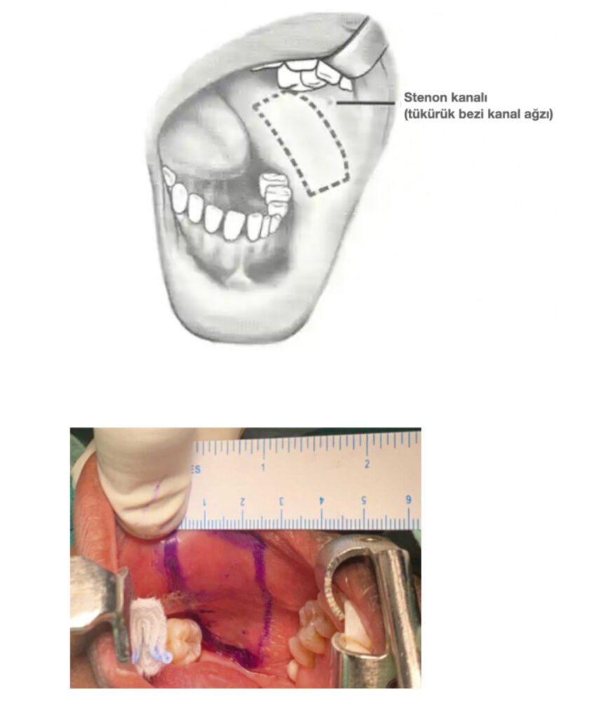
Robotic Ureteroplasty with Buccal Mucosa has opened a new era in the treatment of ureteral stenoses.

Thanks to the method of robotic surgery applied for the first time in our country, the stenosis was completely treated by suturing a piece of buccal mucosa taken from the mouth to the 8 cm long narrow ureter in the form of a patch using the robotic surgery method. The area of the piece taken from the inner part of the lips is healed in a short time thanks to the self-renewal feature of the oral mucosa. The piece of buccal mucosa taken in the operation belongs to the body itself; therefore, it is not rejected by the body. Buccal mucosa is the mucous membrane that covers the inner part of the mouth in the cheeks or the lips.
It has advantageous features in terms of being the person’s own tissue, being well nourished from the vessels and not forming stones in contact with urine.

Especially for the ureteral stenoses longer than 2-3 cm, ureteroplasty with buccal mucosa transplantation using robotic surgery is currently performed with nearly 100% success rates. In our country, it is successfully applied by Prof. Dr. Tibet Erdoğru, who has a say in advanced robotic surgery.

CLICK HERE to watch robotic surgical ureteroplasty surgery with buccal mucosa transplantation from the inner part of the lips.

Treatment of prostate, bladder, kidney and testicular cancers with robotic and laparoscopic surgery in urology

Our Clinic
Prof. Dr. Tibet Erdoğru – Urology & Robotic Surgery    | Data Privacy  |  Cookie Policy

Copyright © 2023 All Rights Reserved  –
⚕️ Immunitas Digital Agency