
Menu
BLADDER CANCER
Bladder cancer is a formation of malignant tumor cells in the tissues that make up the bladder wall.
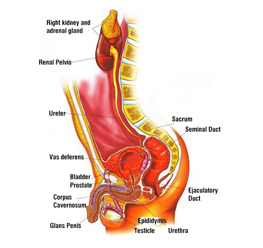
Bladder (urinary bladder, vesica urinaria) is a round shaped organ located in the lower part of abdomen and it collects urine. Bladder, whose wall composes of irregularly arranged and interwoven elastic muscle fibers, resembles a small balloon and thanks to these muscle bundles, it enlarges when filled with urine and gets smaller after urination. Urine produced by kidneys as a result of blood filtering process is drained into bladder through a small tube called ureter. Urine is stored there until urination. When the amount of the urine in the bladder reaches a certain amount, one starts to have a ‘’need to urinate’’, and urine is systematically drained off from bladder through a canal named urethra. (The organs mentioned are shown in Image-1)
Image-1: Connection between kidney, ureter, bladder and the urinary canal called urethra (male anatomy)
According to National Cancer Institute’s estimations, by year of 2022 number of new bladder cancer cases will be 81.180 in USA (In 2010, 70.530 cases were diagnosed annually). It is predicted that 17.100 patients will lose their lives due to bladder cancer in 2022 (Number was 14.680 patients in 2010).
Between 2012 and 2018, for 5 years relative survival rate was 77.1%
What are the types of bladder cancer?
Bladder cancer begins in the cells that line up the inner lining of bladder. 3 main types of cancer (malignant tumors) may develop in bladder.
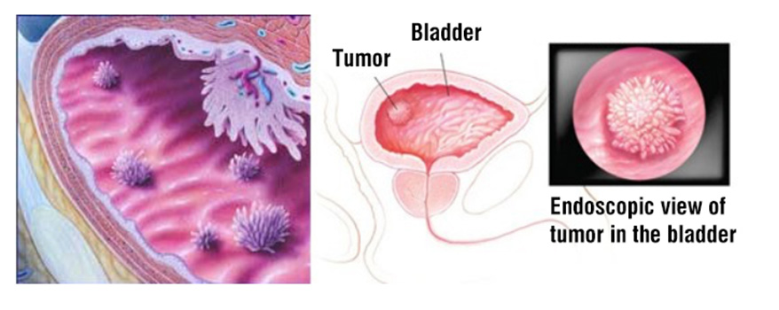
Transitional Epithelial Cell Carcinoma (Urothelial Carcinoma): This type of cancer begins in the cells that line inside of the bladder, which are also called epithelial cells. It is by far the most frequent type of bladder cancers. It accounts for more than 90% of all bladder cancers. It can be either a single papillary tumor, or can be more solid and extended in bladder. (Image-2)
Image-2: Bladder tumors are generally in papillary form as it can be seen in image. They can be either a single tumor, or multifocal tumors in bladder.
Squamous Cell Carcinoma: This type of cancer starts due to squamous cells that develop in bladder following bladder’s exposure to chronic infections and long-term irritations. This type accounts for around 6-8% of all bladder cancers.
Adenocarcinoma: This type of cancers develops from glandular (secretory) cells in the bladder. These secretory glandular cells line the bladder wall and they are responsible for secreting mucus. This type accounts for about 2% of all bladder cancers.
In transitional epithelial cell (urothelial) carcinoma, which is by far the most frequent type of bladder cancers, cancer develops form the epithelial cells lining the inner surface of the bladder. If the cancer is limited only to the epithelial cells that line the inner surface of the bladder and has not spread into the muscle bundles, this is called non-invasive bladder cancer.
Cancer progression generally occurs when it spreads into the muscle layer that lies below these epithelial cells. After invading the muscle layer, this cancer formation reaches the outer layer of the bladder wall and spreads into the fatty tissue around the bladder. This is called invasive bladder cancer. And this process is followed by cancer invasion into lymph nodes through lymphs, and to the other parts of the body from there.
Bladder cancer occurs more often in men than women, more often in White individuals.
Most significant risk factors for developing bladder cancer are tobacco especially smoking cigarettes, gender and diet.
Conditions that increase the likelihood of developing a disease are called risk factors.
Risk factors that increase the possibility of developing bladder cancer are respectively listed below:
Smoking
Exposure to chemicals used in textile, dye and tire industry, with no precautions
Overconsumption of fried and fatty foods
Advanced age, white race, males
Parasitic urinary tract infections with Schistosoma haematobium that especially affect bladder
Other risk factors for bladder cancer include
Having a family history of bladder cancer
Having certain changes in the genes that are linked to bladder cancer, for example RB1, PTEN/ MMAC1, HRAS, NAT2 and GSTM1.
Taking the Chinese herb Aristolochia fangchi
Drinking water from a well that has high levels of arsenic
Drinking water that has been treated with chlorine
Using urinary catheters for a long time
What may be the clinical symptoms of bladder cancer?
First and most common symptom we come across in patients with bladder cancer is blood in the urine or painful urination. Reason of these and other signs stated above might be presence of bladder cancer. However, some other diseases might have these signs, too. So, when someone experiences these kind of symptoms, he/she should visit a doctor.
Blood in urine (color of the urine can be light red and cloudy, in the color of weak tea or wastewater of washed meat.
Very frequent need to urinate
Pain or burning during urination
Pain in the lower tummy, where the bladder is located
Tests and procedures used to diagnose bladder cancer:
Physical Examination
Urinalysis (Complete Urine Analysis)
Urine Cytology: In this test urine sample of the patient is examined under a microscope in order to detect presence of abnormal cells.
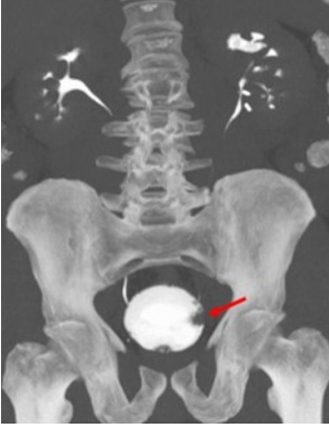
Intravenous Pyelogram (IVP): Evaluation of the urinary collecting system, the urine canal called ureter and bladder using a contrast material injected Intravenously that fills these parts of the body, which is later removed by kidneys.
Image-3: View of the tumor formations in bladder provided by IVP evaluation (red arrow)
Evaluation of Bladder Cancer
Computed Tomography (CT): Along with providing detailed evaluation in terms of cancer presence in kidney and bladder before and after the contrast material injection, it also gives an idea about whether the cancer has spread into surrounding tissues (Image-4). On the other side, recently PET-CT has also been used to determine cancer stage of our patients (Image-5).
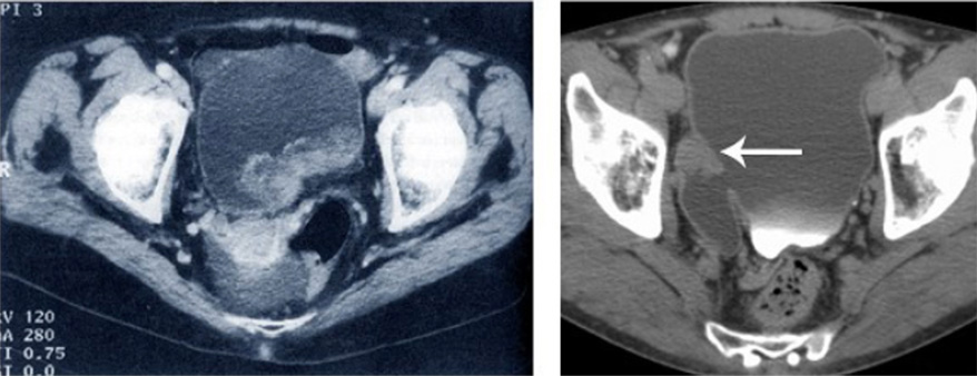
Image-4: On the left, CT image of a rather large tumor formation, which has spread into the fatty tissue on the outer layer of the bladder wall. On the right, you can see the CT image of a bladder diverticulum and tumor’s connection with it.
Bladder tumor and its formation
Image-5: Computed tomography and PET-CT images of bladder tumor
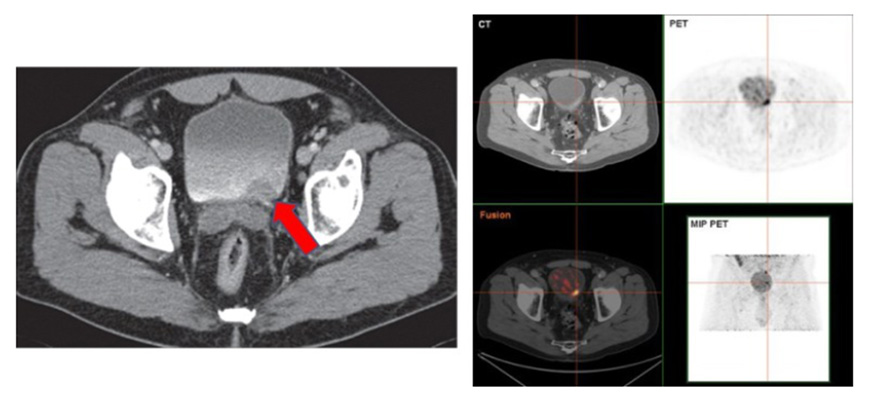
Red arrow: Bladder tumor
Ct image of bladder tumor
Magnetic Imaging: MRI techniques does not have any additional advantages over CT scanning in terms of bladder cancer detection. However, thanks to the newly developed iron-oxide based and diffusion MRI techniques, making a detailed investigation can be possible in order to identify the staging of bladder cancer and see whether there is especially a lymphatic spread or not (Image-6 and Image-7).
Image-6: View of the mass that causes a filling defect on bladder sidewall (pointed with red arrow) and MRI image of bladder tumor
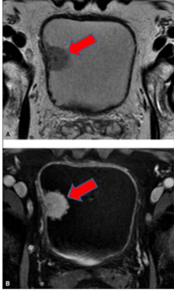
MR image of the bladder tumor
Image-7: In the Diffusion-MRI scan which is able to show lymph node spreads, activity involvement of lymph nodes in the area of iliac artery and venous blood vessel pointed with red arrows may show spread of bladder cancer cells into lymph nodes.
Spread of bladder cancer
Cystoscopy: It is an evaluation procedure performed by inserting a long and thin telescope which has light and optical system on its end through urethra and visualize the inside of the bladder directly. While female patients will not need any form of anesthesia, in men, cystoscopy is carried easily with local anesthesia obtained by an anesthetic applied to the urethra. During this procedure, biopsy samples may be taken with small biopsy instruments from areas that are suspicious for tumor presence. Final diagnosis can be established thanks to pathologic evaluation of these biopsy samples. However, we receive valuable information about the bladder tumor with images provided by cystoscopy, too.
Image-8: Intravesical evaluation using cystoscopy (female patient) / Evaluation of bladder tumor
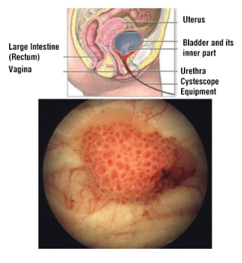
Bladder Tumor
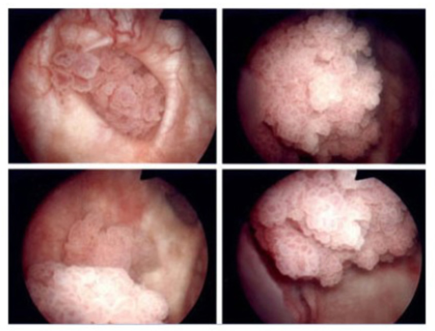
Image-9: Cystoscopy image of cancerous tissue in bladder (looks like bunch of grapes)
Image of bladder tumor
Factors affecting treatment options and disease prognosis (likelihood of recovery after the treatment)
All the possibilities affecting treatment and recovery possibilities
Stages of cancer: Whether the cancer is invasive or non-invasive is the most important prognostic factor as it has been mentioned above. With appropriate treatment, chances of cure are very high in bladder cancers diagnosed at early stages.
Prognosis in Bladder Cancer
The likely outcome or course of a cancer called as “prognosis”
If urinary bladder cancer has been diagnosed on you, you may have many questions about how it could be serious cancer and your chances survival. The likely outcome or course of a disease called “prognosis”
The prognosis of bladder cancer depends on
The stage of the cancer
– has not reached through the inner lining of bladder wall, called as non-invasive superficial cancer
– has spread through the inner lining of the bladder and into the muscle wall of the bladder or beyond it, called as muscle invasive bladder cancer or invasive bladder cancer..
The type of bladder cancer
Whether the cancer is low grade and high grade
The patient’s age and general health care
For non-muscle invasive superficial bladder cancer, prognosis also depends on whether
There are many tumors more than 4 or large tumors more than 4cm diameter
The cancer has grown into the connective tissue next to the lining of the bladder
The cancer has come back after treament such as intravesical BCG or Mitomycine-C instillation.
Superficial non-invasive blazer cancer can often be cured.
For muscle invasive cancer, prognosis also depends on if carcinoma in situ also present and solid formation of the tumor.
5 year survival rate of the bladder cancer is 77%
The 5-year relative survival rates of bladder cancer as as follows,
96% for carcinoma in situ of the bladder alone
77% for localized bladder cancer (cancer is in the bladder only)
39% for regional bladder cancer (cancer has spread beyond the bladder to nearby lymph nodes)
8% for metastatic bladder cancer
Treatment of prostate, bladder, kidney and testicular cancers with robotic and laparoscopic surgery in urology
Our Clinic

- Hacı adil cad. Zerrin sokak, No:2/2 Levent, 34330 Beşiktaş İstanbul Turkey
Prof. Dr. Tibet Erdoğru – Urology & Robotic Surgery | Data Privacy | Cookie Policy