Prof. Dr. Tibet ErdoğruAdvanced Robotic Surgery
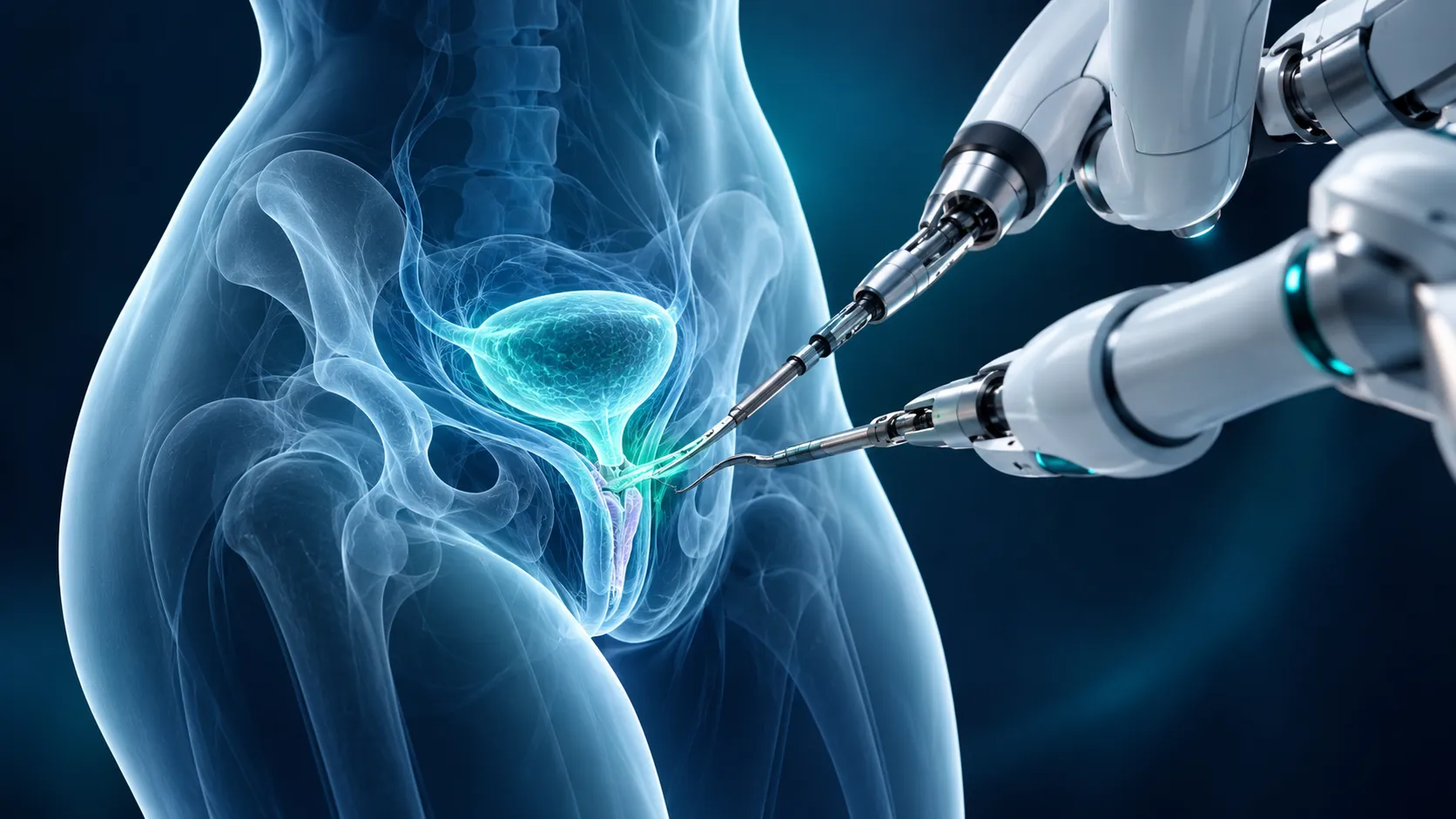
Illustration of robotic surgical arms at the bladder and vagina
Female urology · Robotic reconstruction

VVF: Women's Nightmare: Robotic Repair of Vesicovaginal FistulaProf. Dr. Tibet Erdoğru - Advanced Robotic Surgery

Vesicovaginal fistula is the continuous leakage of urine from the vagina caused by an opening between the bladder and the vagina, most often seen in women after injury during gynaecological surgery. Robotic surgery gives us a very important advantage in the successful repair of vesicovaginal fistula.

Prof. Dr. Tibet Erdoğru
Urology & Robotic Surgery
Repair with robotic surgeryMore than 15 years of robotic surgery experienceIndividual surgical assessment
01 / Understanding the fistula

It may be the cause of
constant urine leakage.

Vesicovaginal
fistula (VVF)An abnormal connection between the bladder and the vagina
Information is the first step towards taking a more informed part in your treatment.

What is a fistula?

A fistula is a passage or channel that forms between two hollow organs in your body (for example, a vesicovaginal fistula is a channel between the bladder and the vagina) or between a hollow organ and your skin (for example, a vesicocutaneous fistula is a channel between the bladder and the skin).

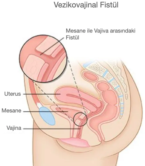
Vesicovaginal fistula (VVF) is an abnormal connection between the bladder and the vagina in women; it causes continuous, uncontrollable leakage of urine from the vagina, known as "total incontinence".

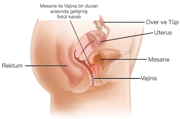
Vesicovaginal fistula: anatomical drawing showing the fistula between the bladder and the vagina
Figure 1: Vesicovaginal fistula developing between the anterior wall of the vagina and the posterior wall of the bladder.
02 / Causes and symptoms

Why does VVF develop?

The main symptom is involuntary leakage of urine from the vagina; this leads to skin irritation and recurrent urinary tract infections.

01 — DIFFICULT CHILDBIRTH

Tissue damage
during childbirth

The most common cause of VVF is tissue death (necrosis): during a difficult delivery, the baby's head presses the mother's soft tissues against the pelvic bone for a long time, and the tissues under pressure no longer receive enough blood supply. Necrosis of the tissues results in a fistula between the bladder and the vagina.

02 — PELVIC SURGERY

After
hysterectomy

In countries with developed obstetric care, VVF is usually an unwanted (iatrogenic) complication of pelvic surgery, especially hysterectomy (removal of the uterus – seen in one of every 1,800 hysterectomies).

03 — RADIOTHERAPY

After
radiotherapy

In addition, vesicovaginal fistula can develop when radiation impairs the blood supply of the tissues during radiotherapy for pelvic cancers.

iThe cause, location and size of the fistula determine the treatment method. The assessment is therefore individual.
03 / Assessment

How is vesicovaginal
fistula assessed?

01

Pelvic examination

Performed to identify the fistula tract on the vaginal side. If the fistula is small or located deep/posteriorly, it is detected by injecting methylene blue into the bladder and seeing the tampon placed in the vagina turn blue.

02

Cystoscopy

If there is a risk of ureterovaginal fistula, it is performed together with retrograde pyelo-ureterography.

03

Voiding cystography

Used to determine the location, presence and diameter of the fistula.

04

Abdominal MRI or CT scan

Performed particularly to rule out injury to the ureter and to look for other pelvic conditions accompanying the problem.

Anatomical drawing showing a fistula tract between the bladder and the anterior vaginal wall
Figure 2: The fistula between the bladder and the vagina may reach 3–4 cm in diameter, and several fistula tracts may have formed.
04 / Repair with robotic surgery

What you should know about robotic VVF repair

When the VVF is large or does not respond to conservative (non-surgical) approaches such as long-term urinary catheterisation, surgical reconstruction – that is, closure – is required. For fistulas caused by surgical injury, the success rate reported for open surgery is generally around 76–97%. Vesicovaginal fistula repair consists of completely removing the fibrotic fistula tract and closing the bladder and vaginal walls separately. Different surgical techniques are recommended depending on the cause, location and size of the fistula and on the surgeon's experience.

Although the vaginal approach is less traumatic for the patient (lower morbidity/side effects) than open abdominal repair (and is a more familiar method, especially for gynaecologists), the abdominal approach is generally preferred in the following situations:

  • Large (>2 cm in diameter) or supratrigonal (above the bladder trigone) fistulas.
  • Fistulas located very close to the ureteral orifices.
  • Especially patients who have had failed or multiple complex transvaginal/transabdominal VVF repairs.

Regardless of all this, for robotic surgeons with advanced experience in reconstructive robotic surgery, repair with robotic surgery is the most effective method for reducing the morbidity of the abdominal approach.

Whatever the size of the fistula, whatever its cause, and even after previous failed repairs, robotic surgery in our experience is associated with a complete success rate, minimal surgical trauma and lower morbidity, and allows the patient to recover faster.
Specific indications for the robotic approach

01

Insufficient field of view due to a narrow vagina or a high-lying or retracted fistula.

02

The fistula being very close to the ureter.

03

An accompanying pelvic condition such as ureteral injury (ureterovaginal fistula), or a complex fistula involving another intra-abdominal organ.

04

The presence of more than one fistula.

05

Cases with a previous failed transvaginal or open abdominal repair attempt.

06

Vesicovaginal fistulas that developed after radiotherapy.

05 / Step by step

The steps of robotic vesicovaginal fistula repair

The images are from a real operation. Surgical photos stay blurred until you choose to open them.

01

Protecting the ureters

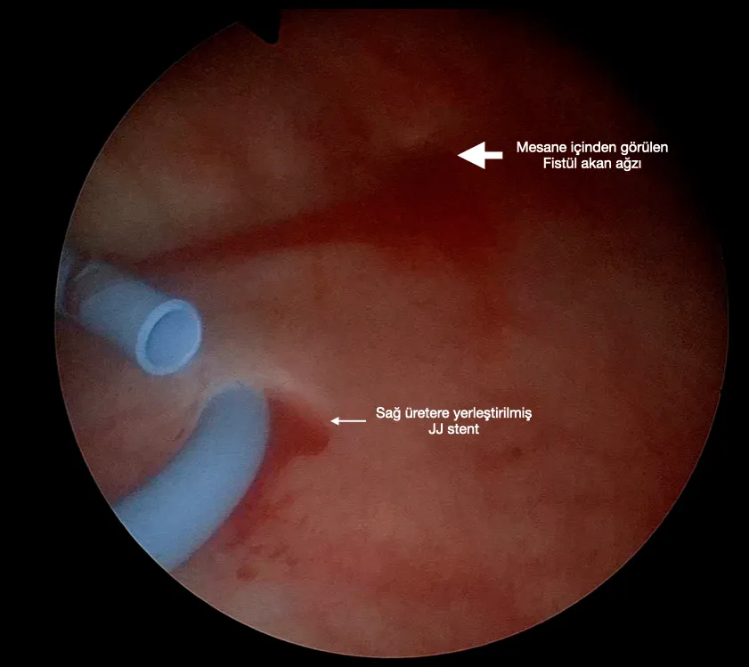
If there is a risk of ureterovaginal fistula, retrograde pyelography is performed with cystoscopy and a JJ stent is placed in both ureters. This ensures that the ureters are identified and protected while the fistula tract is removed and the walls are closed (Figure 3).

Cystoscopic view: JJ stent in the right ureter and the fistula opening
Figure 3: Cystoscopic view of the right ureteral orifice with a catheter (JJ stent) and the fistula line very close to it.
02

Separating the scar tissue

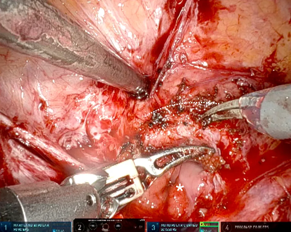
The dense scar tissue that has formed between the bladder and the vagina due to previous operations is separated by sharp dissection after the bladder is filled with 60–90 ml of saline. Figure 4 shows, in a highly complex fistula case, the vaginal opening caused by dense scar tissue (white star) and severe adhesions (white dashed line).

Robotic surgery image: separating scar tissue between the vagina and the bladder
Figure 4: Severe scar tissue between the anterior vaginal wall and the posterior bladder wall (white dotted line).
03

Removing the fistula tract

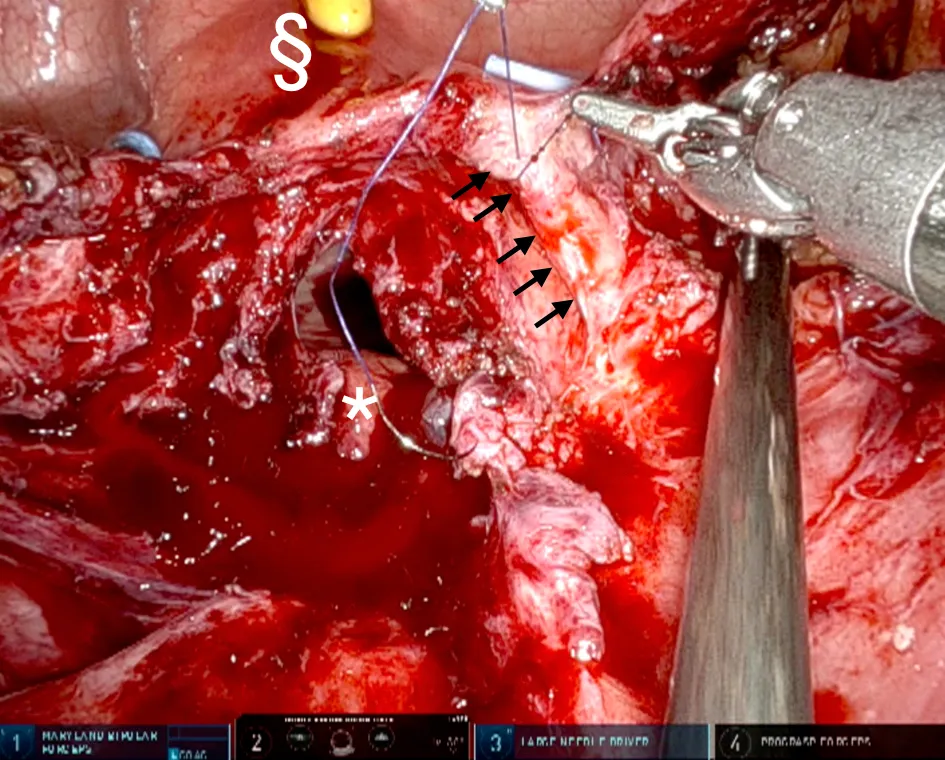
The dense scar tissue around the fistula and the fistula tract are dissected until the healthy bladder wall and vaginal wall are completely exposed. All scar and fistula tract tissue is removed; healthy tissue must be identified for a higher success rate. Especially in patients with a history of traumatic surgery, a ureterovaginal fistula (a fistula tract running directly from the ureter to the vagina) may be found and repaired separately with 5-0 Vicryl sutures (Figure 5).

All steps of the robotic surgery in the case shown in these photos can be watched on video on Prof. Dr. Tibet Erdoğru's YouTube channel.

Robotic surgery image: repair of the right ureteral fistula site
Figure 5: Right ureteral fistula site repaired with 5-0 Vicryl (black arrows); §: bladder opened in the midline, with bilateral JJ stents and the Foley catheter visible inside.
04

Two-layer closure of the vaginal wall

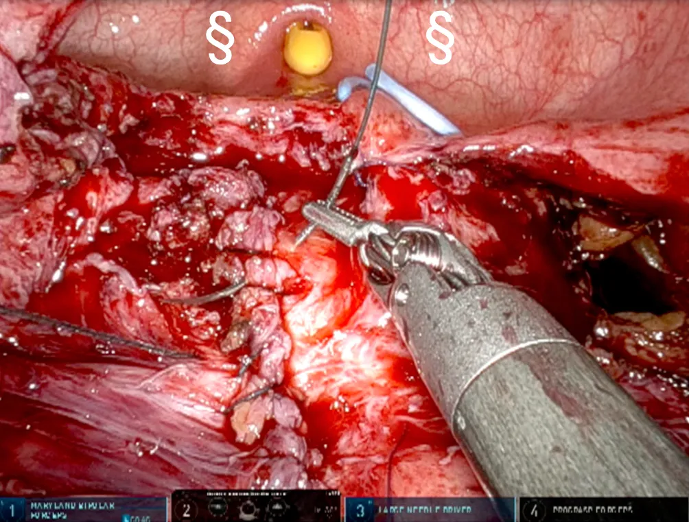
With the high-resolution image of robotic surgery, magnified 15 times compared with the naked eye, the fistula lying deep in the body and the unhealthy tissue around it are removed; the anterior vaginal wall is then closed tightly with V-Loc sutures, leaving no gap at all, as shown in Figure 6a. In addition, as seen in Figure 6b, a second layer of reinforcement and closure must always be added, again using 3-0 V-Loc sutures.

Robotic surgery image: first suture layer of the vaginal wall
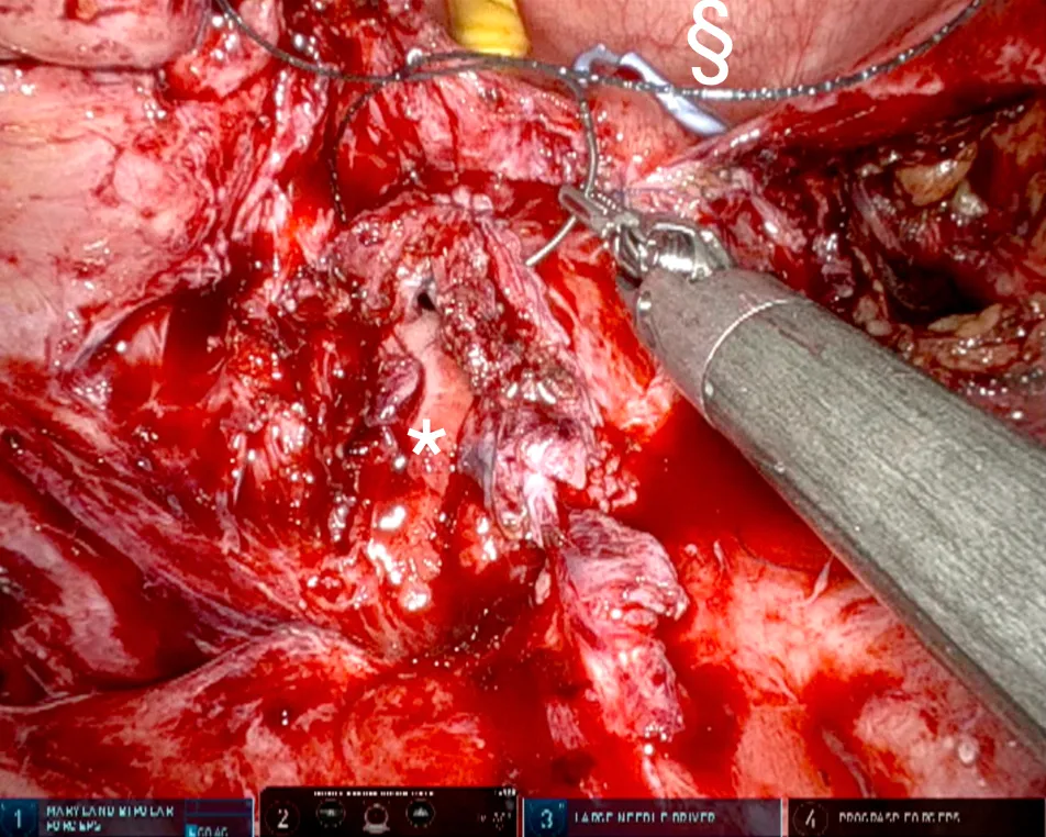
Figure 6a: The anterior vaginal wall closed with a first layer of continuous V-Loc sutures. * Inside the vagina, § inside the bladder.
Robotic surgery image: second suture layer of the vaginal wall
Figure 6b: The anterior vaginal wall closed with a second, securing layer of V-Loc sutures. § Inside the bladder.
05

Protective omental flap

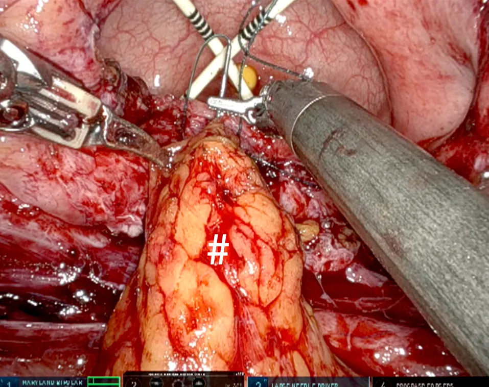
Before or after the midline bladder opening is closed, an omental flap is placed in one or two layers between the repaired vagina and the bladder, both to act as a barrier in the resected area and to secure blood supply through the vessels it contains (Figure 7). After the flap is fixed over the vaginal repair line, the ureteral stents are replaced with single-J 7 Fr catheters and the opened bladder wall is closed with 3-0 V-Loc sutures.

Robotic surgery image: omental flap placed between the vagina and the bladder
Figure 7: Protective omental layer placed between the vagina and the bladder.
06 / Video

Watch robotic VVF repair

Press the play button to start the video.
Better information, more informed decisions.

Discover more videos on urology and robotic surgery.

Explore the YouTube channel
07 / After surgery

Care and follow-up
after surgery.

Surgical care after robotic VVF repair is no different from open surgery. The only difference is that recovery is faster and there is less pain after robotic surgery.

Drain monitoring

The drain is removed 1 or 2 days after surgery, when the daily drainage falls below 50 ml. The patient is discharged with one Foley bladder catheter and two single-J ureteral catheters.

Ureteral catheters

In our experience, placing single-J ureteral stents is very helpful, because it keeps the bladder almost completely dry, which supports the healing of the vaginal and bladder walls. These catheters therefore stay in place for 7 days.

Foley catheter

The urethral Foley catheter is removed between days 14 and 21 after surgery, depending on the quality of the reconstruction and the surgeon's preference. Cystography is not always necessary.

Advice for patients

Patients are advised not to use intravaginal tampons and to avoid sexual intercourse for 2 months.

Antibiotics

Prophylactic (preventive) antibiotics are usually given after surgery until all catheters are removed. Any urinary tract infection that develops after the urethral catheter is removed must be treated.

Contact & appointments

Let's assess your
treatment together.

To learn more about vesicovaginal fistula and its repair with robotic surgery, or to book an appointment, please contact our clinic.

Call for an appointment Message us on WhatsApp
CLINIC PHONE+90 549 804 03 08PROF. DR. TIBET ERDOĞRU CLINIC
Levent Mahallesi, Hacı Adil Caddesi, Zerrin Sokak
No: 2/2, 34330 İç Levent, Beşiktaş / İstanbul
View on map ↗Contact page ↗
WhatsApp
Prof. Dr. Tibet Erdoğru
Contact & appointments
Call the clinic ↗WhatsApp